Procedure Details
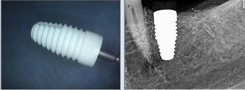
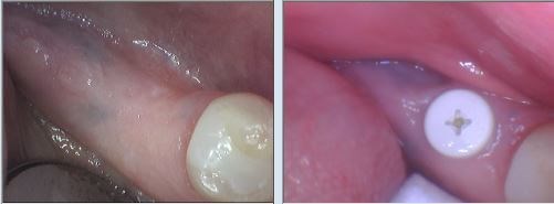
The patient presented with a missing tooth on the lower left. She was worried about her bite and ability to chew since she would need other future extractions. Her goal was to have an implant placed, however she had an allergy to metal and wasn't sure it would be possible. A CBCT (Cone Beam CT) was taken in our office and revealed that the bone structure and space made her a good candidate for implant placement. Due to her metal allergy, a Zirconia implant was placed. Dr. Patel continues to monitor her healing and the patient will return for the implant abutment and crown.
Procedure Details
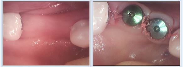
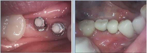
The patient presented with two missing teeth on the lower right. She was having trouble chewing and wanted to replace them with implants. A CBCT (Cone Beam Catscan) was taken in our office, which revealed there was bone loss in the area, however the remaining bone and space made her a good candidate for implants. Dr. Patel placed bone grafts in each area to help increase the bone density, resulting in the stability needed. Once the implants were placed Dr. Patel continue to monitor the patient's healing and approximately two months later he restored the implants with abutments and crowns. The patient continues with her routine visits in our office along with her own homecare to maintain good oral health.
Procedure Details
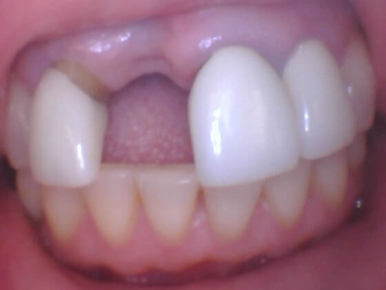
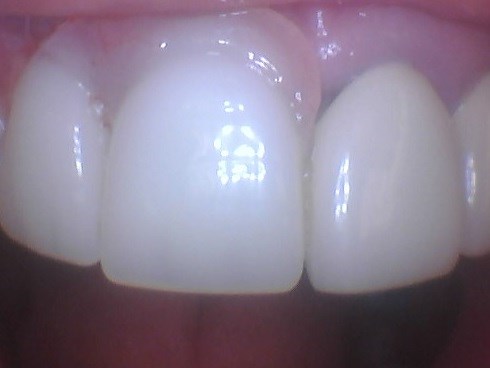
The patient presented for a second opinion due to her history of recent dental treatment that generated complications. These included infection and bone loss around a recently placed implant, as well as recession of the gums around it and adjacent teeth. With the aid of digital diagnostic tests such as an intra-oral camera, digital x-rays and cone beam ct scan Dr. Patel verified infection and un-restorability of the adjacent tooth. After discussing benefits and alternatives with the patient, Dr. Patel helped with removal of the infected tooth, that would have compromised the longevity of the recently placed implant. The cone beam ct scan revealed bone loss around the implant, but that it could be restored. For the purpose of maintaining space, providing a cosmetic solution and helping the bone graft healing, a transitional partial was provided at the end of surgery for the healing period. The patient was monitored with follow up appointments and in 2-6 months she will be ready to restore her implant.
Procedure Details
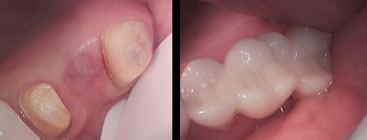
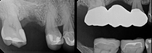
The patient presented with a broken tooth and missing crown. A digital x-ray and CBCT (cone beam catscan) were taken. They revealed a fracture within the tooth, deep decay and an active infection. Dr. Patel diagnosed the tooth as unrestorable and recommended an extraction. Over the next several weeks, the patient was seen for follow up appointments to monitor healing of the extraction site. To replace the tooth, the patient chose to have a bridge placed, as the adjacent teeth had also presented with decay and required crowns. Using our Waterlase laser, Dr. Patel was able to access the...

