Procedure Details
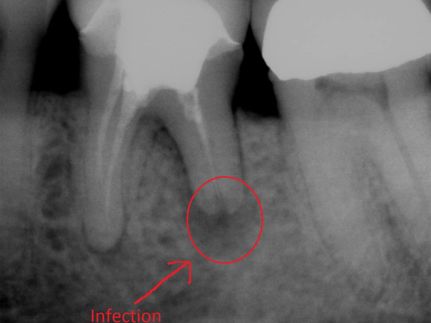
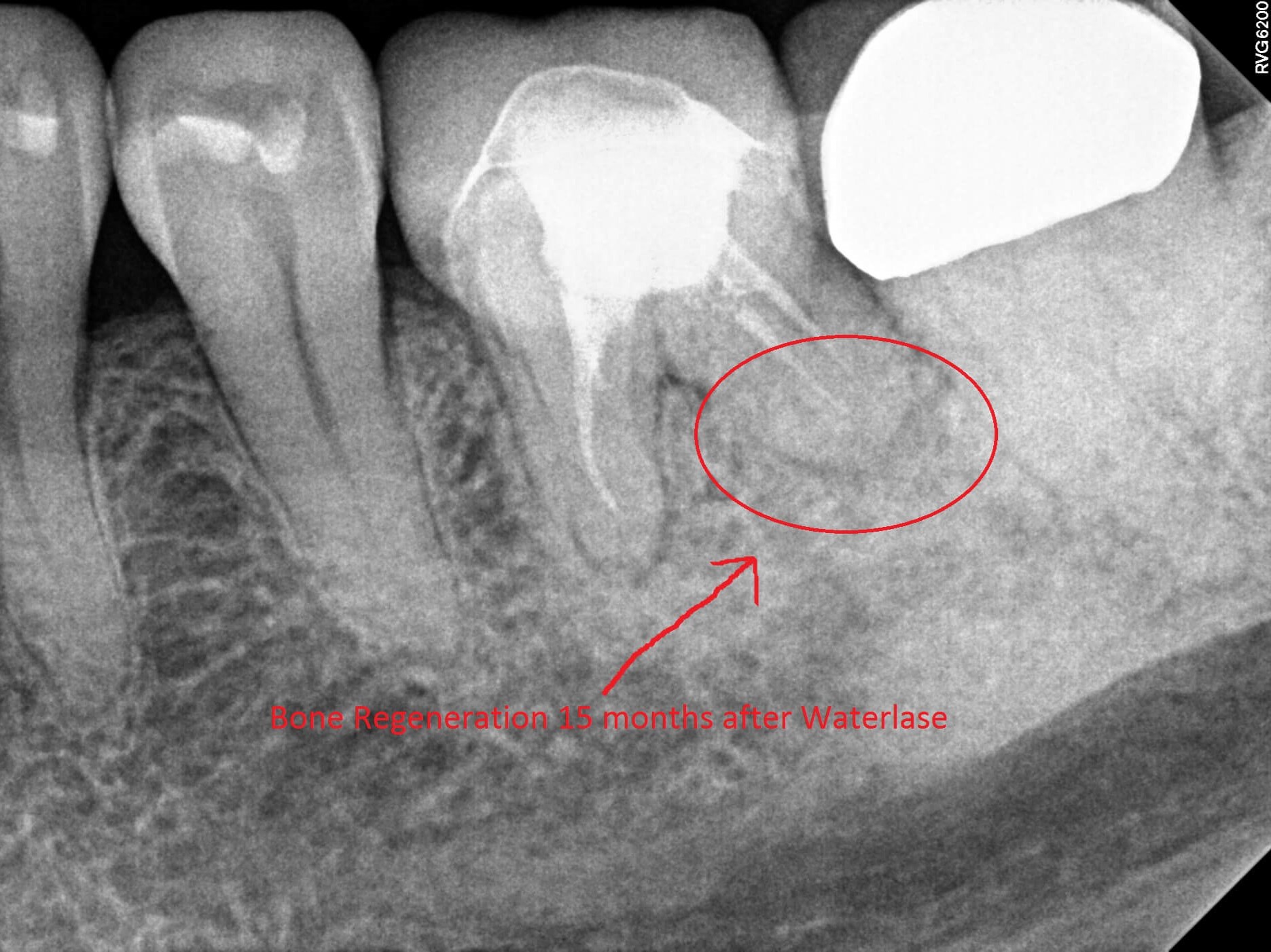
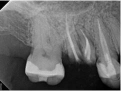
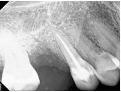
The patient presented for an emergency appointment to address pain. The tooth previously had a root canal completed, so there should not have been a feeling of pain in the actual tooth. A digital x-ray was taken and showed a PARL (Periapical Radiolucency, a change indicating bacteria and infection at the tip of the tooth root.) A periodontal (gum) and endodontic abscess was diagnosed as well as minimal bone loss. An incision and drainage along with antibiotics were recommended as treatment. Using the Waterlase laser, Dr. Patel accessed the infection, cleaned out the bacteria and diseased tissue below the gum line and around the bone. We continued to monitor the patient at follow up appointments. Another digital x-ray, shown here, was taken 15 months later and bone regeneration was noted. She was able to continue with her routine care in our office.


Procedure Details
The patient was seen in our office for a dental emergency. She was experiencing pain and sensitivity when biting and chewing. The pain woke her up in the middle of the night, prompting her to request an emergency appointment. An exam and xray revealed that a tooth on her lower right was abscessed and due to bone loss, was now non restorable. Dr. Patel diagnosed that the tooth needed to be extracted. After discussing replacement options, the patient decided that she wanted to replace the tooth with an implant. The tooth was extracted and due to insufficient bone structure, a bone graft was placed in the site. A few months later once the site healed, an implant was placed and subsequent appointments were made to check the healing and implant stability. The implant was restored about three months later with a same day crown. (Depending on the patient's healing time and regeneration of bone, the entire implant process usually takes between 5-12 months.) Once her treatment was completed, the patient was instructed to continue with her routine care in our office.

Procedure Details
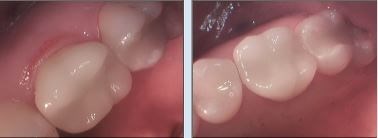
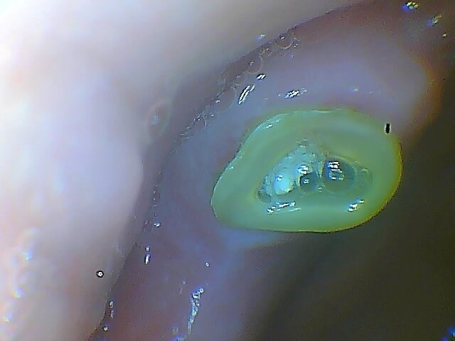
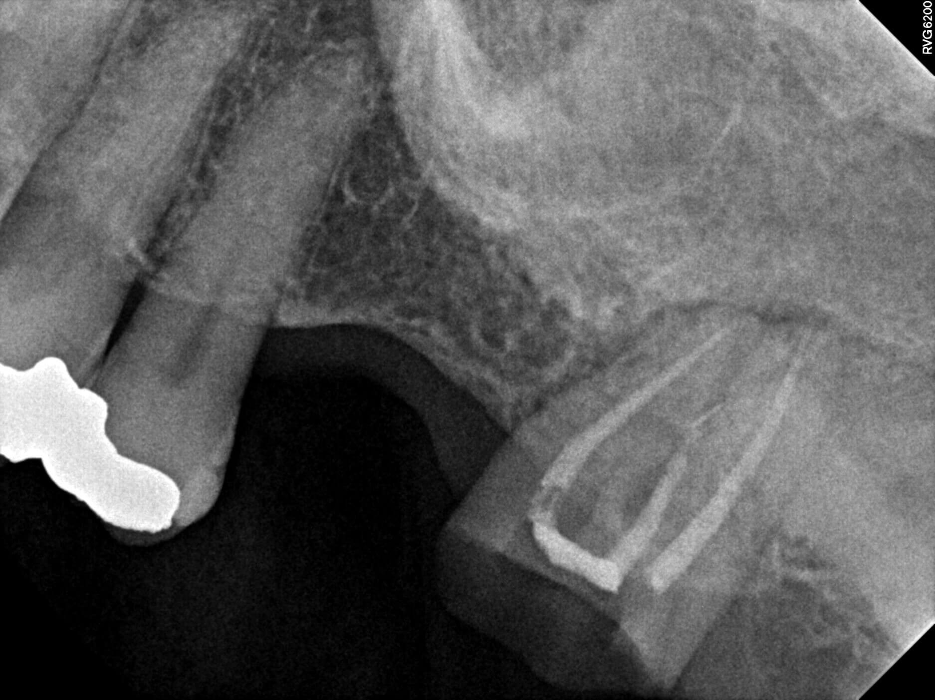
The patient presented with concern about an old silver filling that had begun to chip away on her upper left. She was embarrassed about the look and condition of the tooth. During her exam, a digital x-ray revealed recurrent decay under the existing amalgam filling and cracks within the tooth. The old filling was removed and the tooth was excavated with the help of our WaterLase laser to access the subgingival decay. Due to the extensive decay, Dr. Patel recommended that a pulp cap be placed to help protect the unexposed nerve tissue. The tooth structure was built up for support and a crown was milled using our same day CEREC chamber. Once her occlusion was checked and any necessary adjustments were made for a correct fit and bite, the crown was permanently cemented.
Procedure Details
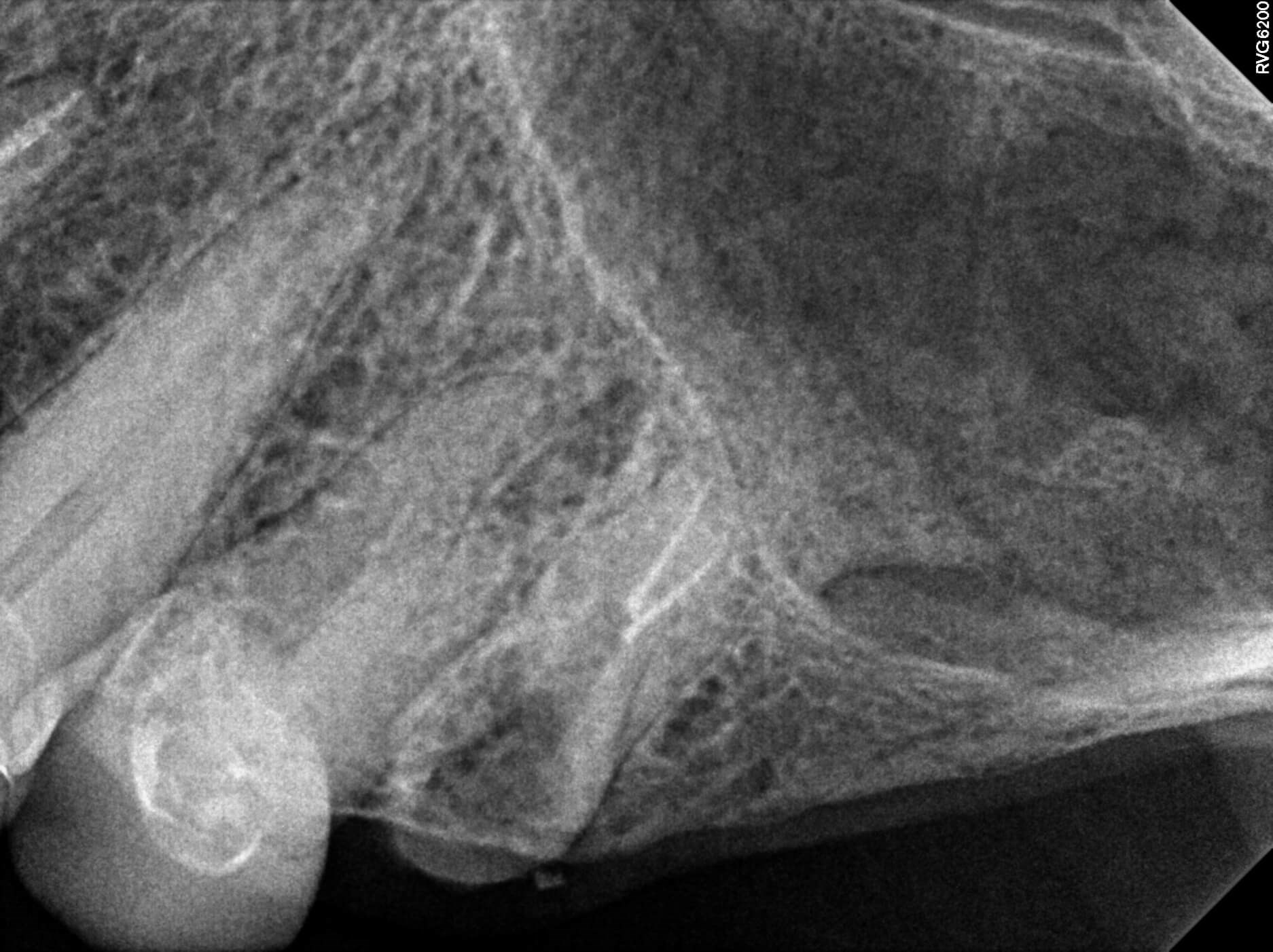
The patient presented for an emergency appointment for a broken tooth. He arrived in "excruciating pain" and was unable to eat or sleep. The tooth had broken off at the gumline and a digital radiograph revealed that an infection was present. Due to the active infection and condition of the remaining tooth structure, Dr. Patel diagnosed it as unrestorable and recommended an extraction. He was able to remove the tooth the same day and prescribed antibiotics to help clear any remaining infection. The patient's healing was monitored at a follow up appointment and he was able to resume his normal hygiene maintenance.
Procedure Details
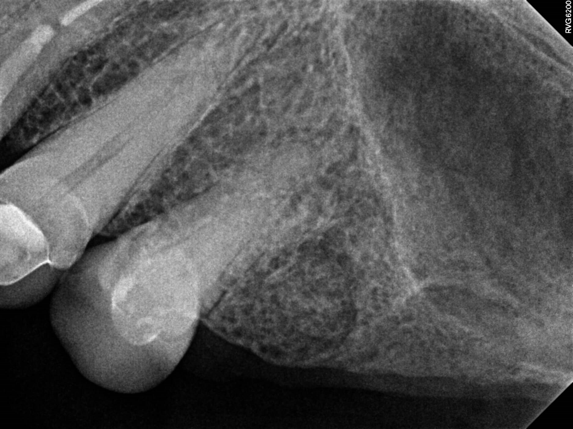
This patient was seen for an emergency appointment to address pain and a loose crown. A digital x-ray revealed there was decay present and the tooth was cracked. Due to the mobility and condition of the tooth, Dr. Patel recommended an extraction. To relieve the patient's pain, the extraction was performed during his emergency visit. Dr. Patel monitored the patient's healing and he was able to continue his routine hygiene appointments in our office.
Procedure Details
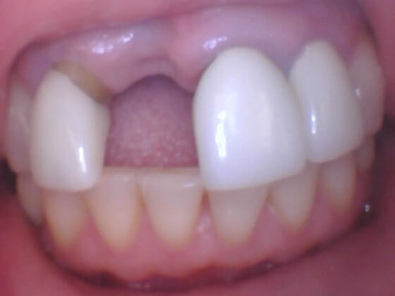
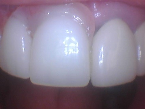
The patient presented for a second opinion due to her history of recent dental treatment that generated complications. These included infection and bone loss around a recently placed implant, as well as recession of the gums around it and adjacent teeth. With the aid of digital diagnostic tests such as an intra-oral camera, digital x-rays and cone beam ct scan Dr. Patel verified infection and un-restorability of the adjacent tooth. After discussing benefits and alternatives with the patient, Dr. Patel helped with removal of the infected tooth, that would have compromised the longevity of the recently placed implant. The cone beam ct scan revealed bone loss around the implant, but that it could be restored. For the purpose of maintaining space, providing a cosmetic solution and helping the bone graft healing, a transitional partial was provided at the end of surgery for the healing period. The patient was monitored with follow up appointments and in 2-6 months she will be ready to restore her implant.


Procedure Details
The patient presented with broken teeth and an esthetic concern. A digital x-ray revealed a deep depth of decay in the patient's last three lower left teeth. Intraoral photos were taken and reviewed with the patient in order for him to visually see the need for treatment. Dr. Patel recommended composite tooth colored fillings. The teeth were excavated with laser assistance to access decay below the gum line. Composite tooth-colored fillings were placed, his occlusion was checked and bite adjusted. The patient will return for his routine hygiene appointments.
Procedure Details
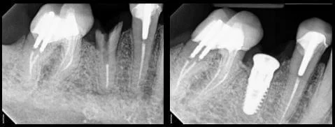
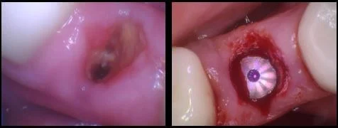
The patient presented for an emergency appointment because he had broken a tooth on the lower right. Dr. Patel examined the patient and reviewed his x-ray, which revealed the tooth was fractured and un-restorable. He recommended an extraction and an implant as replacement. After the tooth was extracted, Dr. Patel placed a bone graft, due to there being insufficient bone structure for an implant. Since the bone graft provided good primary stability, the patient was able to receive his implant the same day. Dr. Patel will monitor the patient's healing and in a couple months will be able to restore the implant with a CEREC crown.
Procedure Details
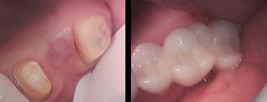
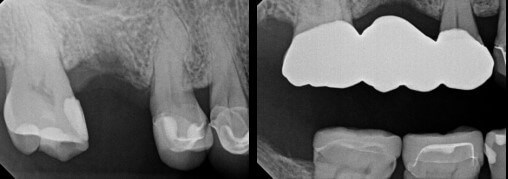
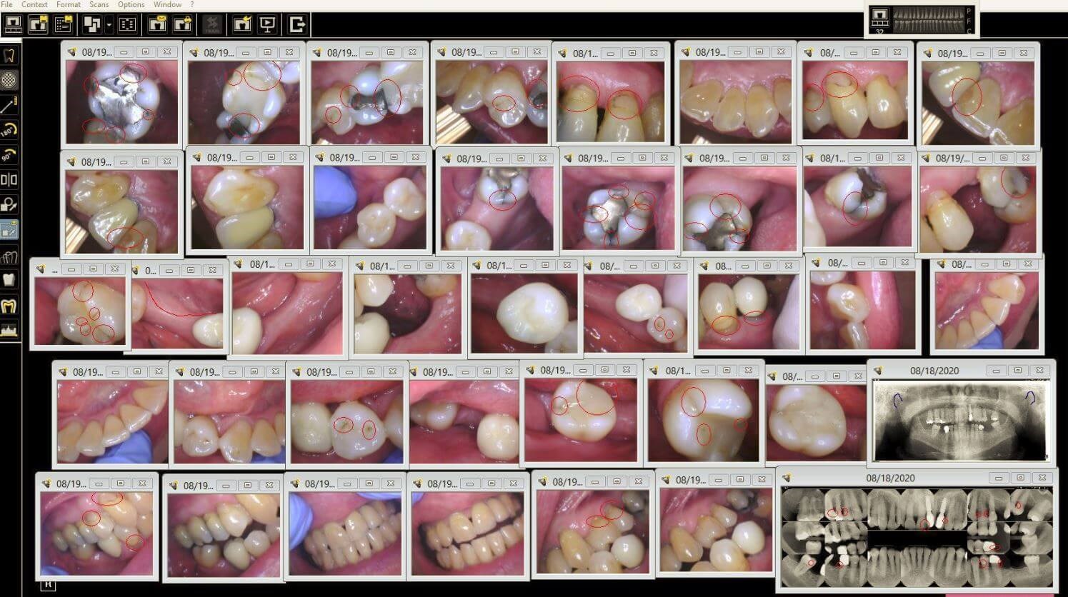
The patient presented with a broken tooth and missing crown. A digital x-ray and CBCT (cone beam catscan) were taken. They revealed a fracture within the tooth, deep decay and an active infection. Dr. Patel diagnosed the tooth as unrestorable and recommended an extraction. Over the next several weeks, the patient was seen for follow up appointments to monitor healing of the extraction site. To replace the tooth, the patient chose to have a bridge placed, as the adjacent teeth had also presented with decay and required crowns. Using our Waterlase laser, Dr. Patel was able to access the...
Procedure Details
During your initial visit, we will review your medical and dental history, as well as gather any medical clearances if necessary. You will be able to discuss what motivates you to seek dental care and any emotional concerns or fears you may have. We will collect diagnostic information including but not limited to x-rays and intraoral photos. Dr. Patel will perform an oral cancer screening, examine your gum tissue and evaluate the condition of your jaw joints and bite. We will chart any prior or existing decay and review oral hygiene instructions with you. Once all of the diagnostic information has been gathered, you will be scheduled back for a full treatment plan consultation with Dr. Patel.
Procedure Details
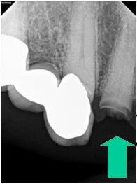
The patient presented with a cracked crown on an upper right tooth. An emergency exam and digital x-ray revealed deep decay and a fractured root, resulting in the tooth being un-restorable. Due to insufficient bone in the area and the patient's desire to eventually replace the missing tooth, Dr. Patel recommended an extraction and bone graft to help support a future implant. The patient was seen for follow up visits to monitor his healing and eventually began the implant process.
Procedure Details
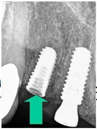
The patient presented for an emergency appointment to address a tooth on the upper left that had broken off at the gum line. During an exam with Dr. Patel, a digital x-ray revealed the tooth had decay into the nerve and was not salvageable. An extraction was recommended, along with a bone graft, due to insufficient remaining bone structure to support an implant as replacement. The patient's healing was monitored and two months later the implant was placed. Dr. Patel continued to monitor the patient's progress and one month later, the implant was stable with zero mobility noted. A same day CEREC crown was provided as the final implant restoration.


Procedure Details
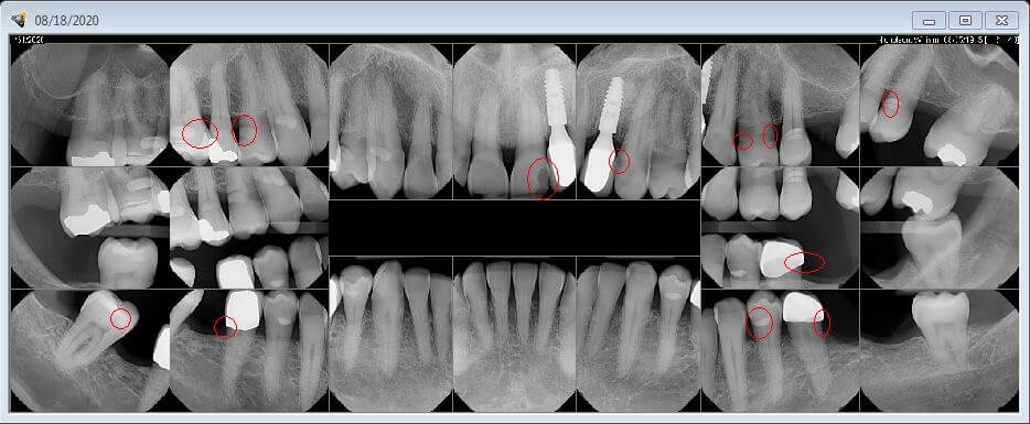
The patient presented for a second opinion after seeing several other dentists, regarding her concern of decay. She had staining and "black spots" she could see on her teeth along with an old amalgam (silver) filling that she felt was falling apart. During an exam with Dr. Patel, digital x-rays were taken, as well as photos using our intraoral camera. The x-rays were examined using our Logicon technology and revealed active decay present. Dr. Patel diagnosed a plan to remove the old filing and decay. The teeth were excavated and due to the depth of decay, a protective liner was placed in each tooth to help calm the nerves and prevent root canals. Composite (tooth colored material) was filled and shaped in each tooth. The patient will continue to be seen for her routine cleanings and exams to help ensure the health of her teeth and gums.


