
Procedure Details
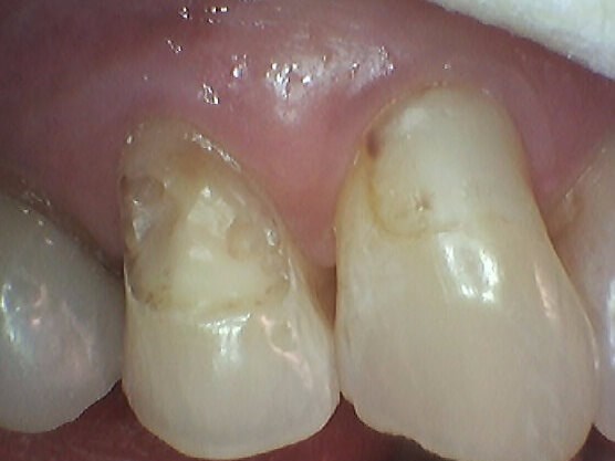
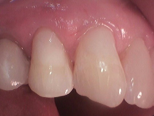
The patient presented with broken teeth and an esthetic concern. A digital x-ray revealed a deep depth of decay in the patient's last three lower left teeth. Intraoral photos were taken and reviewed with the patient in order for him to visually see the need for treatment. Dr. Patel recommended composite tooth colored fillings. The teeth were excavated with laser assistance to access decay below the gum line. Composite tooth-colored fillings were placed, his occlusion was checked and bite adjusted. The patient will return for his routine hygiene appointments.
Procedure Details
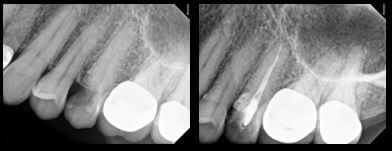
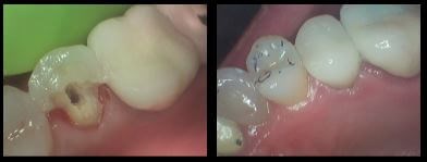
The patient presented for an emergency visit due to a broken tooth. During an exam with Dr. Patel, digital x-rays revealed a vertical crack, requiring a root canal. The tooth was excavated and evaluated, while an intraoral camera was used to record diagnostic photos of the treatment process. The canals were cleaned and disinfected with the help of our Biolase laser and then filled with a material called "gutta percha". Dr. Patel placed a temporary filling at the access until the patient returned a week later for her permanent crown. The temporary filling was taken out and once all decay was removed, the remaining tooth structure was insufficient to support a crown. A post was placed and the tooth was built up to provide stability. Using our CEREC computer and milling chamber, Dr. Patel designed a crown that was then cemented the same day.
Procedure Details
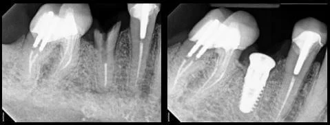
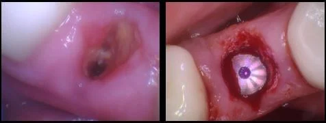
The patient presented for an emergency appointment because he had broken a tooth on the lower right. Dr. Patel examined the patient and reviewed his x-ray, which revealed the tooth was fractured and un-restorable. He recommended an extraction and an implant as replacement. After the tooth was extracted, Dr. Patel placed a bone graft, due to there being insufficient bone structure for an implant. Since the bone graft provided good primary stability, the patient was able to receive his implant the same day. Dr. Patel will monitor the patient's healing and in a couple months will be able to restore the implant with a CEREC crown.
Procedure Details
During your initial visit, we will review your medical and dental history, as well as gather any medical clearances if necessary. You will be able to discuss what motivates you to seek dental care and any emotional concerns or fears you may have. We will collect diagnostic information including but not limited to x-rays and intraoral photos. Dr. Patel will perform an oral cancer screening, examine your gum tissue and evaluate the condition of your jaw joints and bite. We will chart any prior or existing decay and review oral hygiene instructions with you. Once all of the diagnostic information has been gathered, you will be scheduled back for a full treatment plan consultation with Dr. Patel.


Procedure Details
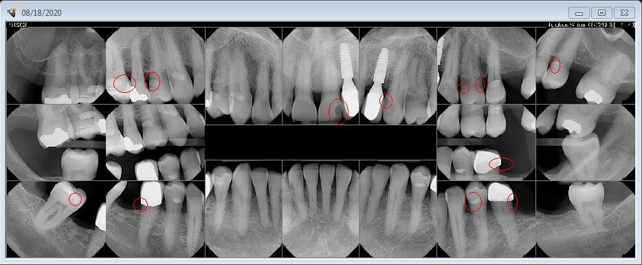
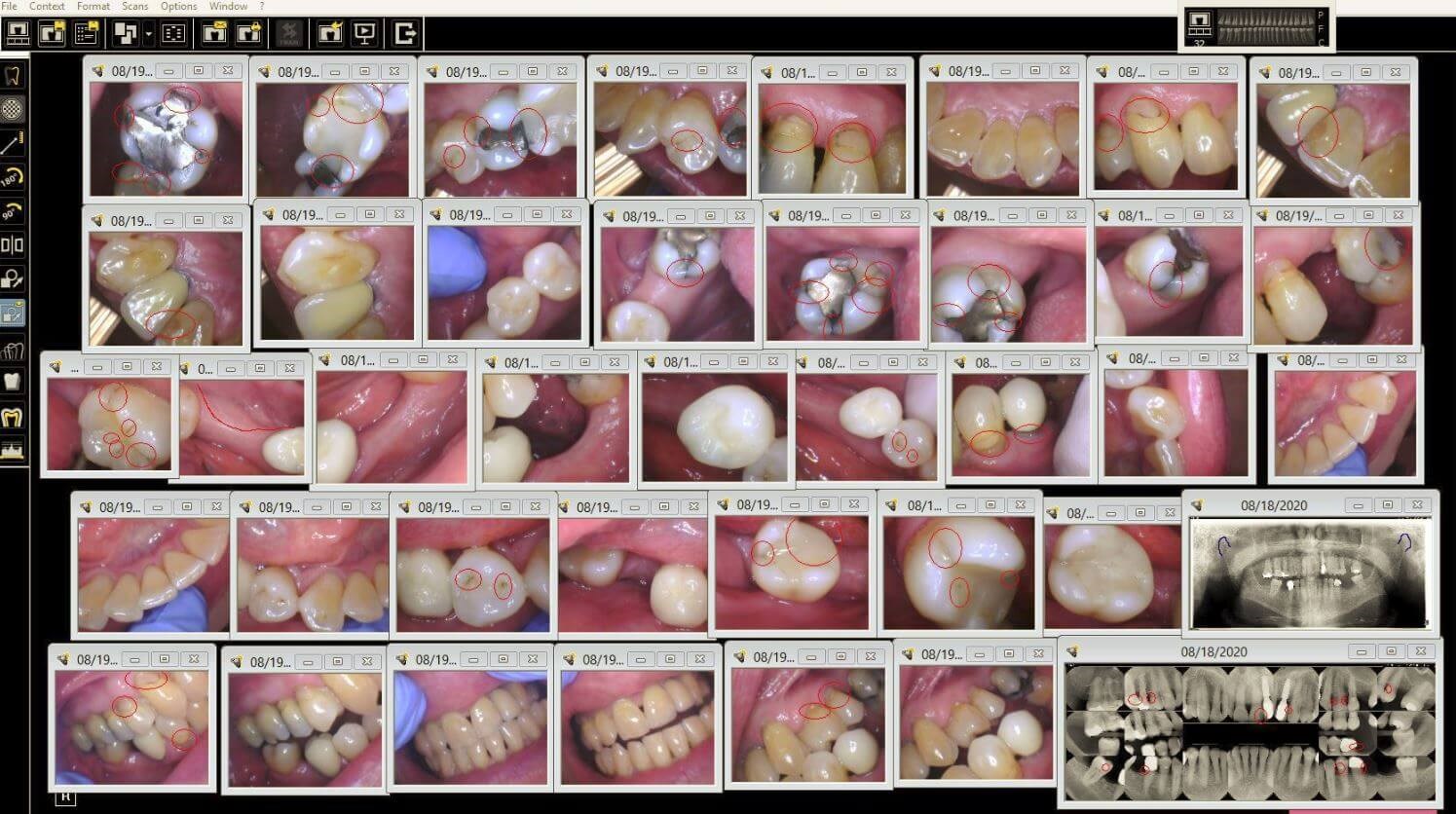
The patient presented for a second opinion after seeing several other dentists, regarding her concern of decay. She had staining and "black spots" she could see on her teeth along with an old amalgam (silver) filling that she felt was falling apart. During an exam with Dr. Patel, digital x-rays were taken, as well as photos using our intraoral camera. The x-rays were examined using our Logicon technology and revealed active decay present. Dr. Patel diagnosed a plan to remove the old filing and decay. The teeth were excavated and due to the depth of decay, a protective liner was placed in each tooth to help calm the nerves and prevent root canals. Composite (tooth colored material) was filled and shaped in each tooth. The patient will continue to be seen for her routine cleanings and exams to help ensure the health of her teeth and gums.


